The anticipation of a new life is a journey filled with wonder, questions, and a deep desire for reassurance. In the modern medical landscape, the most powerful tool for bridging the gap between an expectant mother and her developing baby is Ultrasonography in Pregnancy. Often referred to as the “window to the womb,” this technology has revolutionised prenatal care, allowing clinicians to monitor growth, detect potential issues early, and provide parents with the peace of mind they need.
At Tirsa Healthcare (Tirsa Healthcare), we believe that an informed pregnancy is a healthy pregnancy. This guide explores how ultrasonography works, why it is safe, and the critical milestones it tracks throughout the three trimesters.
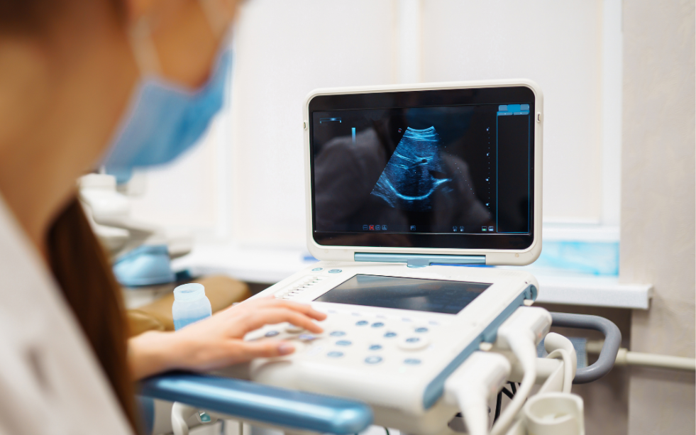
What is Ultrasonography in Pregnancy?
Unlike X-rays, which use ionizing radiation, Ultrasonography in Pregnancy utilizes high-frequency sound waves to create images. A device called a transducer is moved across the abdomen, sending sound waves into the body. These waves bounce off the baby’s tissues, bones, and organs, returning as echoes that a computer translates into a real-time visual image on a monitor.
Because it does not involve radiation, it is considered the gold standard for fetal monitoring. For decades, millions of scans have been performed globally with no documented ill effects on the mother or the developing fetus, provided they are performed by trained medical professionals.
The Timeline: Key Scans and Their Purposes
A standard, healthy pregnancy usually involves several specific ultrasound milestones. Each scan serves a distinct clinical purpose.
1. The Early Pregnancy Scan (6–9 Weeks)
Often, the first scan a woman receives, the primary goal here is “viability.” The sonographer confirms the pregnancy is located within the uterus (ruling out an ectopic pregnancy) and checks for a heartbeat. This scan also determines if you are expecting one baby or multiples.
2. The Dating Scan (11–13 Weeks)
Accuracy is vital in the early stages. By measuring the baby from the top of the head to the buttocks (Crown-Rump Length), doctors can establish a highly accurate due date. During this window, the Nuchal Translucency (NT) Scan is also performed. This measures the fluid-filled space at the back of the baby’s neck, which acts as a crucial screening marker for chromosomal conditions like Down Syndrome.
3. The TIFFA or Anomaly Scan (18–22 Weeks)
This is perhaps the most significant aspect of Ultrasonography in Pregnancy. The Target Imaging for Fetal Anomalies (TIFFA) scan is an exhaustive, head-to-toe examination. The radiologist looks at the structure of the brain, the chambers of the heart, the kidneys, the spine, and the limbs. It ensures that the internal organs are developing correctly and checks the position of the placenta.
4. Growth and Wellbeing Scans (28–36 Weeks)
In the third trimester, scans focus on the “environment” and growth trajectory. The doctor measures the baby’s head circumference, abdominal circumference, and femur length to estimate weight. They also assess the volume of amniotic fluid—the “cushion” of water surrounding the baby—to ensure the baby has enough room and nutrients to thrive until delivery.
Safety First: Dispelling Common Myths
Despite its long history of safety, many expectant parents have concerns about the frequency of scans. It is important to remember:
- Thermal Index: Modern ultrasound machines are designed with “ALARA” (As Low As Reasonably Achievable) principles. They use the minimum amount of energy required to get a clear image.
- Medical Necessity: While it is tempting to want a scan every week to see the baby, Ultrasonography in Pregnancy should always be performed for medical reasons under the guidance of a healthcare provider.
- 3D/4D Scans: These provide lifelike images of the baby’s face and movements. While wonderful for bonding, they should be conducted at certified centers to ensure safety protocols are followed.
Why Technology Matters in Prenatal Care
The quality of the ultrasound image depends on two factors: the sophistication of the machine and the skill of the radiologist. High-resolution equipment can pick up minute details that older machines might miss, such as small septal defects in the heart or subtle markers in the brain.
At Tirsa Healthcare, we emphasise the importance of diagnostic centres that utilise state-of-the-art technology. A clear, accurate scan reduces the need for repeat procedures and provides a definitive roadmap for your obstetrician to follow, and highly skilled radiologist , Dr Nikhil
Preparing for Your Ultrasound
To get the best results from your Ultrasonography in Pregnancy, consider these simple tips:
- Hydration: For early scans (first trimester), a full bladder helps “lift” the uterus, making the baby easier to see.
- Clothing: Wear two-piece outfits (like a top and trousers/skirt) so that only the abdominal area needs to be exposed.
- Questions: Don’t be afraid to ask your sonographer what you are looking at. Seeing the spine, the beating heart, or a waving hand can be a powerful bonding experience.
Conclusion: Partnering with Tirsa Healthcare
The journey to motherhood is a path of transformation. Utilising Ultrasonography in Pregnancy is not just about clinical data; it’s about the first connection you make with your child. It enables early intervention, informed decision-making, and the joy of witnessing growth in real time.
At Tirsa Healthcare , we are committed to connecting you with the finest diagnostic facilities and maternal care experts. We ensure that your prenatal journey is supported by accurate imaging, expert interpretation, and a compassionate approach to your family’s needs.
Hello , Are you ready to schedule your next prenatal milestone? Visit Tirsa Healthcare today to find the most trusted ultrasound services and ensure your baby’s growth is monitored with the precision it deserves.
Recent Comments